Institut universitaire de cardiologie et de pneumologie de Québec, Québec, QC, Canada
paul.poirier@criucpq.ulaval.ca

Introduction
Key Points
- The improvements in risk factor recognition and management that have occurred over the years in modern cardiology may be counteracted by the rising incidence of obesity.
- The presence of obesity may limit the accuracy of the physical exam. It also has the potential to affect the electrocardiogram. Moreover, transthoracic echocardiography can be technically difficult in obese patients, and obese individuals may have several limitations in the catheterization laboratory.
- The obesity paradox may reflect the failure of body mass index to adequately discriminate body fat distribution.
- From a clinical standpoint, both body mass index and waist circumference should probably be assessed and may be useful to better define "at risk" obesity.
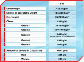
Obesity is a complex multifactorial chronic disorder that develops from an interaction of genotype and the environment. The health hazards of obesity have been recognized for centuries. Populations in industrialized countries are becoming more overweight as a result of changes in lifestyle. Both overweight and obesity must be regarded as serious medical problems in our time since obesity is associated with reduced life expectancy. Indeed, obesity represents an independent predictor of cardiovascular disease (CVD) and this association is more pronounced in individuals under 50 years of age. This is why the American Heart Association stated ten years ago that obesity is a major modifiable risk factor for heart disease [1]. Nowadays, obesity has reached epidemic proportions in the United States as well as in much of the industrialized world and is increasing in prevalence in the developing world [2]. In the most widely used classification of body mass, body weight is expressed in terms of body mass index (BMI) [2]. In adults, obesity is defined as a BMI ≥30 kg/m2, which is further subdivided into grades (Table 1).
The improvements in risk factor recognition and management that have occurred over the years in modern cardiology may be counteracted by the rising incidence of obesity [3]. Beyond an unfavourable risk factor profile, overweight/obesity also affects heart structure and function [2]. However, obesity is a remarkably heterogeneous condition, where the distribution of adipose tissue is of importance in determining the presence/absence of metabolic dysfunctions [4]. Obesity as defined by BMI is undoubtedly associated with an increased rate of comorbidities and cardiovascular mortality [5, 6], and obese individuals considered "at risk" are mostly characterized by features associated with abdominal obesity.
Physical Exam
 [Click to enlarge]
The presence of obesity may limit the accuracy of the physical exam. Jugular venous pulse is often not seen, and heart sounds are usually distant. A common finding in massive obesity is pedal edema, which can occur in part as a consequence of elevated ventricular filling pressure, despite elevation in cardiac output. Obese individuals can also have increased demand for ventilation and breathing workload, especially in the supine position. Accurate blood pressure measurement is crucial since many obese patients are hypertensive. A small cuff size can cause considerable increases in blood pressure. This could incorrectly classify up to 35% of normotensive obese individuals as hypertensive. One should always evaluate the presence of cor pulmonale when examining an obese individual. In obese patients, the split S2, when either inaudible or very poorly defined in the second interspace, is often best heard at the first left interspace. Therefore, an increase in the intensity of P2, suggestive of pulmonary hypertension, may be missed at the bedside.
Surface Electrocardiogram (ECG)
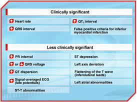
Obesity has the potential to affect the ECG in several ways: 1) displacement of the heart by elevating the diaphragm in the supine position, 2) increasing the cardiac workload and 3) increasing the distance between the heart and the recording electrodes. The voltage of the QRS complexes is attenuated by its passage through a fatladen chest wall and is related to several factors, including the anatomy of the thorax, the degree of fatty infiltration of the heart, the degree of associated chronic lung disease, the increase in left ventricular muscle mass and, most importantly, the selection of the electrocardiographic leads for measuring voltage. Overall, the effect of weight loss in obese patients on the QRS voltage is a source of controversy in the literature; studies report a decrease, no change or an increase in the QRS amplitude after weight reduction. Heart rate, PR interval, QRS interval, QRS voltage and QTc interval all showed an increase with increasing obesity. An increased incidence of false-positive criteria for inferior myocardial infarction was reported in both obese individuals and in women in the final trimester of pregnancy, presumably because of diaphragmatic elevation (Table 2).
Echocardiography
 [Click to enlarge]
 [Click to enlarge]
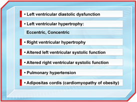
Transthoracic echocardiography can be technically difficult in obese patients, and obtaining a good echocardiographic window is often difficult. This is of importance when evaluating the presence of left ventricular diastolic dysfunction. Pulmonary venous Doppler evaluation may be used but if it is not technically feasible, transmitral Doppler imaging with the use of the Valsalva maneuver may properly evaluate the presence of left ventricular diastolic dysfunction. Another feature of the echocardiographic assessment in obese patients is the differentiation between subepicardial adipose tissue and pericardial effusion, which can at times be difficult. Epicardial adipose tissue is known to be a common cause of pseudopericardial effusion, and this adipose tissue depot may cause an underestimation of the amount of pericardial fluid. Another issue is the presence of fat within the heart. Fat can accumulate in a variety of places, but the site of predilection tends to be the interatrial septum. Lipomatous hypertrophy of the interatrial septum should be suspected in the presence of a dumbbell-shaped appearance of the septum, with thick echogenic tissue surrounding a thin echo at the level of the fossa ovalis. Also, accumulation of fat may simulate a mass. Several heart functions can be unmasked with an echocardiogram (Table 3).
Cardiac Catheterization
Obese individuals may have several limitations in the catheterization laboratory. The catheterization laboratory table usually does not accommodate subjects weighing more than 160 kg. Moreover, vascular access to the femoral vein and artery may be difficult. The percutaneous radial approach has advantages in the very obese patient, for whom the percutaneous femoral technique may be technically difficult and bleeding hard to control after catheter removal. Indeed, the frequency of complications using the percutaneous radial technique is very low and should be contemplated when the evaluation of extremely obese individuals is necessary in the catheterization laboratory.
Assessment of Obesity by the Cardiologist
It appears that obesity as defined solely by BMI cannot always discriminate between the individuals at higher risk of developing CVD. Non-obese overweight patients with excess intra-abdominal (visceral) adiposity (i.e., patients therefore at higher risk) may not be detected on the basis of BMI alone [7]. For these reasons, measurement of waist circumference and a set of metabolic markers has been proposed to detect obese individuals at a higher CVD risk [5, 8]. Waist or waist-to-hip ratio (WHR) has been used as a proxy measure for body fat distribution. Abdominal obesity has been reported as a risk factor for CVD worldwide and is likely to better refine clinical assessment of obesity risk [7, 9, 10].
Coronary Artery Disease
Atherosclerosis begins in childhood (5-10 years) and is demonstrated predominantly as fatty streaks. Examination of arteries post-mortem from young individuals (15 to 34 years of age) in the Determinants of Atherosclerosis in Youth (PDAY) study who died from accidental injuries, homicides or suicides revealed that the extent of fatty streaks and even advanced lesions (fibrous plaques and plaques with calcification or ulceration) in the right coronary artery and abdominal aorta were associated with obesity and size of the abdominal panniculus [11]. In adults, it has been shown that 1) maximal density of macrophages/mm2 in atherosclerotic lesions is associated with intra-abdominal obesity [12], 2) reduced coronary flow reserve is related to body fat distribution and insulin resistance [13], 3) the metabolic syndrome is associated with lipid-rich plaque [14], 4) coronary artery calcium and abdominal aortic calcium is associated with intra-abdominal adipose tissue [15]. Prospective evidence shows that abdominal obesity is associated with accelerated progression of carotid atherosclerosis in men independently of overall obesity and other risk factors [16]. Also, the components of the insulin resistance syndrome have been reported, following coronary artery bypass graft, to be associated with angiographic progression of atherosclerosis in non-grafted coronary arteries [17].
The Obesity Paradox
Despite the fact that obesity has been shown to be an independent risk factor for CVD, many studies have reported that obese patients with established CVD have a better prognosis than do patients with ideal body weight: this is the so-called "obesity paradox". This paradox has been best described for patients with advanced systolic heart failure [2] and patients with coronary artery disease [18]. The improved survival of obese individuals is paradoxical principally because of the assumption that excessive weight is always and invariably harmful. As a matter of fact, among patients with congestive heart failure, subjects with higher BMI are at decreased risk for death and hospitalization compared with patients with a "healthy" BMI [2]. Also, obesity was associated, in a prospective cohort study, with lower all-cause and cardiovascular mortality after unstable angina/non-ST-segment elevation myocardial infarction treated with early revascularization [19].
The obesity paradox may reflect the failure of BMI to adequately discriminate body fat distribution [7, 20, 21]. Since BMI measures total body mass, i.e., both fat and lean mass, it may better represent the protective effect of lean body mass on mortality. This negative confounding may have been underappreciated in prior studies that did not adjust for measures of abdominal obesity. It is possible that the favourable prognosis implications associated with mildly elevated BMI might actually reflect the intrinsic limitations of BMI in differentiating adipose tissue from lean mass. BMI's lack of specificity could dilute the adverse effects of excess fat with the beneficial effects of preserved or increased lean mass [22]. As an example, in patients with known CVD or following acute myocardial infarction, overall obesity as assessed by BMI was not associated with myocardial infarction, cardiovascular mortality and total mortality when abdominal obesity (WHR, waist circumference) was integrated into the analysis [9, 23, 24]. Another limitation in most studies reporting an obesity paradox in patients with CVD is that non-intentional weight loss, which would be associated with a poor prognosis, is not assessed, as BMI is measured only at the beginning of the study. Patients who have decompensated heart failure may lose weight because of extensive caloric demands associated with the increased work of breathing, and patients who show poor nutrients absorption by the edematous bowel may be at higher risk of recurrent CVD events.
Despite the high correlation between waist circumference and BMI, the combination of both indices may be very relevant in clinical practice because waist circumference for a given BMI is a strong predictor of all-cause mortality. Studies reporting negative results between all-cause mortality and waist circumference did not mutually adjust for waist circumference and BMI, a possible explanation for the inconsistent results [25, 26]. Another example is that the excess health risk associated with a higher BMI declines with increasing age. An explanation for the lack of a positive association between BMI and mortality at older ages is that, in older persons, higher BMI is a poor measure of body fat and may simply represent a measure of increased physical activity with preserved lean mass. Sarcopenic obesity, which is defined as excess fat with loss of lean body mass, is a highly prevalent problem in older individuals. In fact, the ideal BMI may be higher in older adults than in middle-aged adults. It was recently reported in ~4,000 persons aged ≥75 years that WHR rather than waist circumference predicted mortality in non-smoking men and women, mainly because of the association with cardiovascular deaths [27]. In the Health Professionals Study, in men aged 65 years, waist circumference and WHR were significantly related to CVD mortality [25]. It was found in the Cardiovascular Health Study in over 5,000 patients aged ≥65 years with a mean BMI of 26.3 kg/m2 (42% overweight) that higher BMI values indicated a lower mortality risk once the risk attributable to waist circumference was accounted for, whereas waist circumference values indicated a higher mortality risk once the risk attributable to BMI was accounted for [28]. Death rates were highest in individuals with a high waist circumference within the overweight and obese BMI categories. Finally, in a large case-control study, WHR was found to be more strongly associated than BMI with myocardial infarction, whereas the association with BMI was weak and intermediate for waist circumference in older patients [29]. In order to discriminate low-risk vs. high-risk subjects, WHR could be more useful. Further studies are needed to clarify the concept of the obesity paradox in patients with known CVD.
Assessment of Adiposity in Clinical Practice
The introduction of waist circumference as a simple risk measure in public health settings has already begun, but debate regarding the simplification of the measure is ongoing. Thresholds for waist circumference to identify individuals with excess cardiovascular risk have been suggested, but the choice of waist circumference thresholds should be based on outcomes of importance, such as all-cause mortality or myocardial infarction. It seems from the data available that there is no basis for choosing thresholds because the mortality rate ratio increased steadily with waist circumference. Nevertheless, there may be a difference between different ethnic groups [29]. From a clinical standpoint, both indices should probably be assessed and may be useful to better define "at risk" obesity [11, 30, 31]. It was observed in the IDEA (International Day for the Evaluation of Abdominal obesity) study, which included 157,211 patients, that both waist circumference and BMI were independently associated with the presence of CVD in both men and women [32]. Nevertheless, with all the knowledge available in the literature regarding obesity and CVD, assessment and management of obesity following acute coronary syndrome is simply inadequate [33].
Conclusion
Without a doubt, obesity is a risk factor for CVD. There are numerous clinical indices to evaluate obesity (BMI, waist circumference, WHR). Accurate diagnosis of obesity may lead to more refined assessment of body fat composition/distribution. Over the years, studies have helped refine indices associated with CVD. For example, total cholesterol has been replaced by LDL and HDL cholesterol to better evaluate the patient's risk of CVD. Today, we are no longer using total weight to assess the presence of obesity. Although BMI has been useful in epidemiological studies in order to assess the presence of obesity, it fails to differentiate between differing body compositions. BMI does not characterize excess centrally distributed obesity, which is more consistently associated with adverse effects on metabolism, dyslipidemia and insulin resistance. BMI also can be falsely increased in the presence of increased lean body mass (such as in trained athletes), and low BMI values are associated with chronic conditions leading to loss of lean body mass. Thus, other clinical indices of adiposity such as waist circumference and WHR should be incorporated into the cardiologist's clinical approach in order to better target and manage "at risk" obesity.
References
- Eckel RH and Krauss RM. American Heart Association call to action: obesity as a major risk factor for coronary heart disease. AHA Nutrition Committee. Circulation 1998; 97: 2099-100.
- Poirier P, Giles TD, Bray GA, et al. Obesity and cardiovascular disease: pathophysiology, evaluation, and effect of weight loss: an update of the 1997 American Heart Association Scientific Statement on Obesity and Heart Disease from the Obesity Committee of the Council on Nutrition, Physical Activity, and Metabolism. Circulation 2006; 113: 898-918.
- Olshansky SJ, Passaro DJ, Hershow RC, et al. A potential decline in life expectancy in the United States in the 21st century. N Engl J Med 2005; 352: 1138-45.
- Després JP and Lemieux I. Abdominal obesity and metabolic syndrome. Nature 2006; 444: 881-7.
- Eckel RH, Grundy SM and Zimmet PZ. The metabolic syndrome. Lancet 2005; 365: 1415-28.
- Poirier P and Després JP. Waist circumference, visceral obesity, and cardiovascular risk. J Cardiopulm Rehabil 2003; 23: 161-9.
- Poirier P. Adiposity and cardiovascular disease: are we using the right definition of obesity? Eur Heart J 2007; 28: 2047-8.
- Després JP, Lemieux I, Bergeron J, et al. Abdominal obesity and the metabolic syndrome: contribution to global cardiometabolic risk. Arterioscler Thromb Vasc Biol 2008; 28: 1039-49.
- Poirier P. Recurrent cardiovascular events in contemporary cardiology: obesity patients should not rest in PEACE. Eur Heart J 2006; 27: 1390-1.
- Yusuf S, Hawken S, Ounpuu S, et al. Effect of potentially modifiable risk factors associated with myocardial infarction in 52 countries (the INTERHEART study): case-control study. Lancet 2004; 364: 937-52.
- Zieske AW, Malcom GT and Strong JP. Natural history and risk factors of atherosclerosis in children and youth: the PDAY study. Pediatr Pathol Mol Med 2002; 21: 213-37.
- Kortelainen ML and Sarkioja T. Visceral fat and coronary pathology in male adolescents. Int J Obes Relat Metab Disord 2001; 25: 228-32.
- Kondo I, Mizushige K, Hirao K, et al. Ultrasonographic assessment of coronary flow reserve and abdominal fat in obesity. Ultrasound Med Biol 2001; 27: 1199-205.
- Amano T, Matsubara T, Uetani T, et al. Impact of metabolic syndrome on tissue characteristics of angiographically mild to moderate coronary lesions integrated backscatter intravascular ultrasound study. J Am Coll Cardiol 2007; 49: 1149-56.
- Rosito GA, Massaro JM, Hoffmann U, et al. Pericardial fat, visceral abdominal fat, cardiovascular disease risk factors, and vascular calcification in a community-based sample: the Framingham Heart Study. Circulation 2008; 117: 605-13.
- Lakka TA, Lakka HM, Salonen R, et al. Abdominal obesity is associated with accelerated progression of carotid atherosclerosis in men. Atherosclerosis 2001; 154: 497-504.
- Korpilahti K, Syvanne M, Engblom E, et al. Components of the insulin resistance syndrome are associated with progression of atherosclerosis in non-grafted arteries 5 years after coronary artery bypass surgery. Eur Heart J 1998; 19: 711-9.
- Uretsky S, Messerli FH, Bangalore S, et al. Obesity paradox in patients with hypertension and coronary artery disease. Am J Med 2007; 120: 863-70.
- Buettner HJ, Mueller C, Gick M, et al. The impact of obesity on mortality in UA/non-ST-segment elevation myocardial infarction. Eur Heart J 2007; 28: 1694-701.
- Romero-Corral A, Lopez-Jimenez F, Sierra-Johnson J, et al. Differentiating between body fat and lean masshow should we measure obesity? Nat Clin Pract Endocrinol Metab 2008; 4: 322-3.
- Romero-Corral A, Montori VM, Somers VK, et al. Association of bodyweight with total mortality and with cardiovascular events in coronary artery disease: a systematic review of cohort studies. Lancet 2006; 368: 666-78.
- Romero-Corral A, Somers VK, Sierra-Johnson J, et al. Diagnostic performance of body mass index to detect obesity in patients with coronary artery disease. Eur Heart J 2007; 28: 2087-93.
- Dagenais GR, Yi Q, Mann JF, et al. Prognostic impact of body weight and abdominal obesity in women and men with cardiovascular disease. Am Heart J 2005; 149: 54-60.
- Kragelund C, Hassager C, Hildebrandt P, et al. Impact of obesity on long-term prognosis following acute myocardial infarction. Int J Cardiol 2005; 98: 123-31.
- Baik I, Ascherio A, Rimm EB, et al. Adiposity and mortality in men. Am J Epidemiol 2000; 152: 264-71.
- Visscher TL, Seidell JC, Molarius A, et al. A comparison of body mass index, waist-hip ratio and waist circumference as predictors of all-cause mortality among the elderly: the Rotterdam study. Int J Obes Relat Metab Disord 2001; 25: 1730-5.
- Price GM, Uauy R, Breeze E, et al. Weight, shape, and mortality risk in older persons: elevated waist-hip ratio, not high body mass index, is associated with a greater risk of death. Am J Clin Nutr 2006; 84: 449-60.
- Janssen I, Katzmarzyk PT and Ross R. Body mass index is inversely related to mortality in older people after adjustment for waist circumference. J Am Geriatr Soc 2005; 53: 2112-8.
- Yusuf S, Hawken S, Ounpuu S, et al. Obesity and the risk of myocardial infarction in 27,000 participants from 52 countries: a case-control study. Lancet 2005; 366: 1640-9.
- Poirier P. Healthy lifestyle: even if you are doing everything right, extra weight carries an excess risk of acute coronary events. Circulation 2008; 117: 3057-9.
- Poirier P. Targeting abdominal obesity in cardiology: can we be effective? Can J Cardiol 2008; 24 Suppl D: 13D-7D.
- Balkau B, Deanfield JE, Després JP, et al. International Day for the Evaluation of Abdominal Obesity (IDEA): a study of waist circumference, cardiovascular disease, and diabetes mellitus in 168,000 primary care patients in 63 countries. Circulation 2007; 116: 1942-51.
- Lopez-Jimenez F, Malinski M, Gutt M, et al. Recognition, diagnosis and management of obesity after myocardial infarction. Int J Obes (Lond) 2005; 29: 137-41.


