Influence of Age
Defining CMR - Visceral Adipose Tissue: the Culprit? Causes and Correlates of Visceral ObesityKey Points
- Visceral adiposity increases with age. This is particularly true for men when compared to pre-menopausal women.
- Visceral adipose tissue accumulation with age predicts greater obesity-related health risks.
- Visceral adipose tissue accumulation may partly explain the metabolic deteriorations observed with ageing.
- In clinical practice, a simple approach to identify individuals gaining visceral fat is to measure changes (increases) in waist circumference over time.
Influence of Age on Adipose Tissue Distribution
Several studies have reported that adipose tissue distribution changes with age. Increasing age has been consistently associated with increased visceral adipose tissue deposition in both sexes [1,2]. This section will briefly review the scientific evidence available on this topic.
Several studies have also documented age-related changes in adipose tissue distribution as reflected by an increase in the waist-to-hip ratio, which has been used as an anthropometric index of relative abdominal adiposity [3,4]. In a sample of 52,953 women, it was found that the body weight increase observed with age was more likely to accumulate in the abdominal area than the gluteo-femoral area [3]. In the 1,179 participants of the Baltimore Longitudinal Study of Aging, a waist-to-hip ratio increase was reported across various age groups (from 17 to 96 years of age) in both men and women [5]. In women, the onset of menopause accelerated abdominal fat accumulation. In another sample of Dutch volunteers (men and women), age was found to be a strong correlate of selective abdominal adipose tissue accumulation as estimated by increased waist circumference [6].
In young individuals (men and women) and middle-aged women, excess energy is preferentially stored in subcutaneous fat depots, although visceral adipose stores may also increase [1] selectively in some genetically susceptible individuals [7]. In this regard, the ratio of abdominal subcutaneous to visceral adipose tissue has been shown to be markedly higher in women than in men and to decrease with age in both sexes. In another study, age was again positively correlated to visceral adiposity and negatively correlated with the amount of abdominal subcutaneous adipose tissue [8]. DeNino et al. [9] have even estimated that in non-obese women, visceral adipose tissue increases with age at a rate of 2.36 cm2 per year.
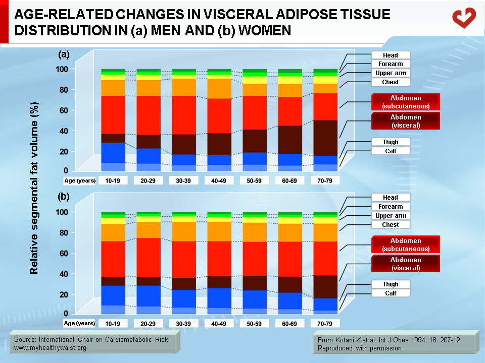
Increased visceral adipose tissue deposition with age is particularly significant among men and post-menopausal women who, on average, have more than twice the amount of visceral adipose tissue than pre-menopausal women [2]. Figure 1 shows the significant increase of visceral adiposity in association with the whole body fat distribution changes with age in men and women. However, even in middle-aged pre-menopausal women, it has been shown that visceral adipose tissue deposition was higher when compared to young women, suggesting that age was associated with an increase in abdominal adiposity in women even before the onset of menopause [10].
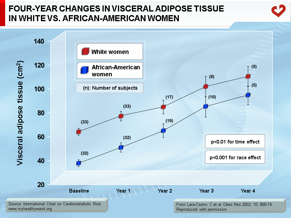
In young pre-menopausal women, age-related changes in visceral adipose tissue relative to total body fat have been reported to be greater than the increase in total body fat over a 4-year follow-up period, and this phenomenon was found to be independent of race (Figure 2) [11]. This study also reported that the age-related increase in total adiposity was greater in African-American women than in white women. However, visceral adiposity was greater in white women at baseline, increased with age, and remained higher in white than in black women.
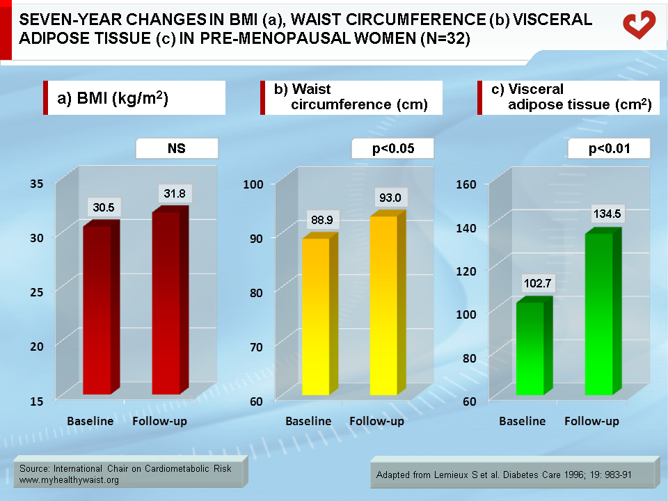
In a 7-year follow-up study of pre-menopausal women, a 30% increase in visceral adiposity was reported although total adiposity indices did not change (Figure 3) [12]. Over the same period, waist circumference, waist-to-hip ratio, and visceral adipose tissue area measured by computed tomography were all significantly increased. In addition, the increase in waist circumference over the 7-year follow-up predicted the increase in visceral adiposity. This latter finding suggest that monitoring changes in waist circumference over time could be useful in clinical practice to detect changes in visceral adiposity with years. This study provides evidence that clinicians should look beyond body weight measurements and the body mass index in order to properly assess whether patients are accumulating dangerous visceral fat over time.
Metabolic Consequences of the Age-related Increase in Visceral Adiposity
It has been clearly established that the prevalence of cardiovascular disease (CVD) increases with age and that the greater number of CVD risk factors in older than younger individuals partly explains this phenomenon [13,14]. For instance, the age-related increase in visceral adiposity has been shown to be an important correlate of alterations in lipoprotein-lipid metabolism and in plasma glucose homeostasis in middle-aged pre-menopausal women when compared to young women [10]. In the same cross-sectional study, when young and middle-aged women with similar levels of visceral adipose tissue and fat mass were compared, some of the well-known age-related differences in the metabolic profile (apolipoprotein B, fasting glucose and glucose area following a 75 g oral glucose challenge) were eliminated. Results from a 7-year follow-up study in pre-menopausal women also showed that the deterioration in plasma glucose and insulin homeostasis indices depended more upon the age-related increase in visceral adipose tissue than upon the increase in total adiposity [12].
Middle-aged men generally have a more atherogenic plasma lipoprotein-lipid profile than young men [15]. However, when middle-aged men with similar total body fat mass and visceral adiposity were compared to young adult men, many age-related differences in the plasma lipoprotein-lipid profile were eliminated, suggesting that the age-related increase in total and visceral adiposity explained many (but not all) age-associated changes in lipoprotein-lipid levels. Lemieux et al. [16] have reported that age is associated with increased LDL particle concentration. The increasing presence of the small, dense LDL phenotype observed in some individuals with age appears to be largely related to an increase in triglyceride levels in both genders [16,17]. As hypertriglyceridemia is another consequence of abdominal obesity—especially visceral obesity—these results highlight the importance of maintaining low levels of visceral fat over time.
Lastly, there is a clear sexual dimorphism regarding changes in abdominal adipose tissue distribution with age, which suggests that abdominal adipose tissue is sensitive to both sex hormones and ageing. Clinicians should be aware of the fact that the increase in visceral adiposity with age predicts a deterioration of their patient’s risk factors profile.
References
-
Enzi G, Gasparo M, Biondetti PR, et al. Subcutaneous and visceral fat distribution according to sex, age, and overweight, evaluated by computed tomography. Am J Clin Nutr 1986; 44: 739-46.
PubMed ID: 3788827
-
Kotani K, Tokunaga K, Fujioka S, et al. Sexual dimorphism of age-related changes in whole-body fat distribution in the obese. Int J Obes 1994; 18: 207-12.
PubMed ID: 8044194
-
Lanska DJ, Lanska MJ, Hartz AJ, et al. Factors influencing anatomic location of fat tissue in 52,953 women. Int J Obes 1985; 9: 29-38.
PubMed ID: 3874840
-
Tonkelaar ID, Seidell JC, van Noord PA, et al. Factors influencing waist/hip ratio in randomly selected pre- and post-menopausal women in the dom-project (preliminary results). Int J Obes 1989; 13: 817-24.
PubMed ID: 2621054
-
Shimokata H, Tobin JD, Muller DC, et al. Studies in the distribution of body fat: I. Effects of age, sex, and obesity. J Gerontol 1989; 44: M66-73.
PubMed ID: 2921472
-
Han TS, McNeill G, Seidell JC, et al. Predicting intra-abdominal fatness from anthropometric measures: the influence of stature. Int J Obes Relat Metab Disord 1997; 21: 587-93.
PubMed ID: 9226490
-
Bouchard C, Tremblay A, Després JP, et al. The response to long-term overfeeding in identical twins. N Engl J Med 1990; 322: 1477-82.
PubMed ID: 2336074
-
Zamboni M, Armellini F, Milani MP, et al. Body fat distribution in pre- and post-menopausal women: metabolic and anthropometric variables and their inter-relationships. Int J Obes Relat Metab Disord 1992; 16: 495-504.
PubMed ID: 1323546
-
DeNino WF, Tchernof A, Dionne IJ, et al. Contribution of abdominal adiposity to age-related differences in insulin sensitivity and plasma lipids in healthy nonobese women. Diabetes Care 2001; 24: 925-32.
PubMed ID: 11347756
-
Pascot A, Lemieux S, Lemieux I, et al. Age-related increase in visceral adipose tissue and body fat and the metabolic risk profile of premenopausal women. Diabetes Care 1999; 22: 1471-8.
PubMed ID: 10480511
-
Lara-Castro C, Weinsier RL, Hunter GR, et al. Visceral adipose tissue in women: longitudinal study of the effects of fat gain, time, and race. Obes Res 2002; 10: 868-74.
PubMed ID: 12226134
-
Lemieux S, Prud’homme D, Nadeau A, et al. Seven-year changes in body fat and visceral adipose tissue in women. Association with indexes of plasma glucose-insulin homeostasis. Diabetes Care 1996; 19: 983-91.
PubMed ID: 8875093
-
Grundy SM, Vega GL and Bilheimer DW. Kinetic mechanisms determining variability in low density lipoprotein levels and use with age. Arteriosclerosis 1985; 5: 623-30.
PubMed ID: 4074195
-
Dedonder-Decoopman E, Fievet-Desreumaux C, Campos E, et al. Plasma levels of VLDL- + LDL-cholesterol, HDL-cholesterol, triglycerides and apoproteins B and A-I in a healthy population– influence of several risk factors. Atherosclerosis 1980; 37: 559-68.
PubMed ID: 7459000
-
Lemieux S, Prud’homme D, Moorjani S, et al. Do elevated levels of abdominal visceral adipose tissue contribute to age-related differences in plasma lipoprotein concentrations in men? Atherosclerosis 1995; 118: 155-64.
PubMed ID: 8579625
-
Lemieux I, Pascot A, Tchernof A, et al. Visceral adipose tissue and low-density lipoprotein particle size in middle-aged versus young men. Metabolism 1999; 48: 1322-7.
PubMed ID: 10535398
-
McNamara JR, Campos H, Ordovas JM, et al. Effect of gender, age, and lipid status on low density lipoprotein subfraction distribution. Results from the Framingham Offspring Study. Arteriosclerosis 1987; 7: 483-90.
PubMed ID: 3675308
 CLOSE
CLOSE
Enzi G, Gasparo M, Biondetti PR, et al. Subcutaneous and visceral fat distribution according to sex, age, and overweight, evaluated by computed tomography. Am J Clin Nutr 1986; 44: 739-46.
PubMed ID: 3788827CLOSE
Kotani K, Tokunaga K, Fujioka S, et al. Sexual dimorphism of age-related changes in whole-body fat distribution in the obese. Int J Obes 1994; 18: 207-12.
PubMed ID: 8044194CLOSE
Lanska DJ, Lanska MJ, Hartz AJ, et al. Factors influencing anatomic location of fat tissue in 52,953 women. Int J Obes 1985; 9: 29-38.
PubMed ID: 3874840CLOSE
Tonkelaar ID, Seidell JC, van Noord PA, et al. Factors influencing waist/hip ratio in randomly selected pre- and post-menopausal women in the dom-project (preliminary results). Int J Obes 1989; 13: 817-24.
PubMed ID: 2621054CLOSE
Shimokata H, Tobin JD, Muller DC, et al. Studies in the distribution of body fat: I. Effects of age, sex, and obesity. J Gerontol 1989; 44: M66-73.
PubMed ID: 2921472CLOSE
Han TS, McNeill G, Seidell JC, et al. Predicting intra-abdominal fatness from anthropometric measures: the influence of stature. Int J Obes Relat Metab Disord 1997; 21: 587-93.
PubMed ID: 9226490CLOSE
Bouchard C, Tremblay A, Després JP, et al. The response to long-term overfeeding in identical twins. N Engl J Med 1990; 322: 1477-82.
PubMed ID: 2336074CLOSE
Zamboni M, Armellini F, Milani MP, et al. Body fat distribution in pre- and post-menopausal women: metabolic and anthropometric variables and their inter-relationships. Int J Obes Relat Metab Disord 1992; 16: 495-504.
PubMed ID: 1323546CLOSE
DeNino WF, Tchernof A, Dionne IJ, et al. Contribution of abdominal adiposity to age-related differences in insulin sensitivity and plasma lipids in healthy nonobese women. Diabetes Care 2001; 24: 925-32.
PubMed ID: 11347756CLOSE
Pascot A, Lemieux S, Lemieux I, et al. Age-related increase in visceral adipose tissue and body fat and the metabolic risk profile of premenopausal women. Diabetes Care 1999; 22: 1471-8.
PubMed ID: 10480511CLOSE
Lara-Castro C, Weinsier RL, Hunter GR, et al. Visceral adipose tissue in women: longitudinal study of the effects of fat gain, time, and race. Obes Res 2002; 10: 868-74.
PubMed ID: 12226134CLOSE
Lemieux S, Prud’homme D, Nadeau A, et al. Seven-year changes in body fat and visceral adipose tissue in women. Association with indexes of plasma glucose-insulin homeostasis. Diabetes Care 1996; 19: 983-91.
PubMed ID: 8875093CLOSE
Grundy SM, Vega GL and Bilheimer DW. Kinetic mechanisms determining variability in low density lipoprotein levels and use with age. Arteriosclerosis 1985; 5: 623-30.
PubMed ID: 4074195CLOSE
Dedonder-Decoopman E, Fievet-Desreumaux C, Campos E, et al. Plasma levels of VLDL- + LDL-cholesterol, HDL-cholesterol, triglycerides and apoproteins B and A-I in a healthy population– influence of several risk factors. Atherosclerosis 1980; 37: 559-68.
PubMed ID: 7459000CLOSE
Lemieux S, Prud’homme D, Moorjani S, et al. Do elevated levels of abdominal visceral adipose tissue contribute to age-related differences in plasma lipoprotein concentrations in men? Atherosclerosis 1995; 118: 155-64.
PubMed ID: 8579625CLOSE
Lemieux I, Pascot A, Tchernof A, et al. Visceral adipose tissue and low-density lipoprotein particle size in middle-aged versus young men. Metabolism 1999; 48: 1322-7.
PubMed ID: 10535398CLOSE
McNamara JR, Campos H, Ordovas JM, et al. Effect of gender, age, and lipid status on low density lipoprotein subfraction distribution. Results from the Framingham Offspring Study. Arteriosclerosis 1987; 7: 483-90.
PubMed ID: 3675308