Other Imaging Techniques
Evaluating CMR - Imaging TechniquesKey Points
- DEXA is a useful tool for assessing total adiposity but is limited in its ability to assess visceral fat.
- Ultrasonography may be a useful tool for assessing visceral fat in clinical practice.
- As measured via ultrasonography, visceral fat thickness is a correlate of cardiometabolic risk factors.
Evaluating CMR: Imaging Techniques
Imaging techniques such as Magnetic Resonance Imaging (MRI) and Computed Tomography (CT) are the gold standard measures for assessing total and visceral fat. However, their cost and limited availability hinder routine widespread use. Other clinical tools, such as dual-energy x-ray absorptiometry (DEXA) and ultrasonography, are useful techniques for assessing total and visceral adiposity and related health risk at a significantly lower cost. The caveat is that these methods are less accurate in measuring visceral fat, which may limit their ability to identify individuals at increased health risk.
Dual-energy X-ray Absorptiometry (DEXA)
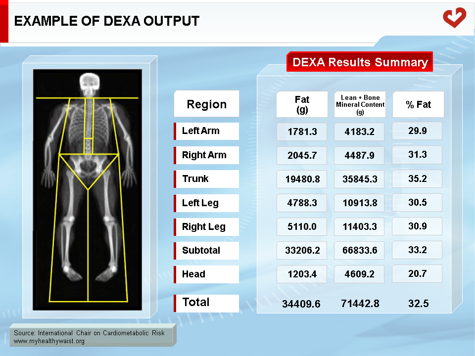
Dual-energy x-ray absorptiometry (DEXA) was originally designed to measure bone mineral content, but it is now commonly used to assess total and regional fat and fat-free mass (Figure 1) [1,2]. DEXA assesses body composition based on the attenuation of x-rays emitted at two energy levels as they traverse the body [1,2]. DEXA provides less radiation exposure than CT and costs significantly less. A whole body DEXA scan requires 15 to 35 minutes depending on the scanner and is relatively easy to use in most populations as it requires very little participant effort [3]. Once the scan is complete, it can be manually subdivided into truncal and appendicular regions. Measures of total and regional lean mass (coefficient of variation=1 to 7%) [4-6] and fat mass (coefficient of variation=1 to 7%) [5] are highly repeatable, but results may differ depending on the scanner model (e.g., Lunar, Holigic, etc.) [7], the software used (algorithms), and the individual’s sagittal diameter and hydration status [3]. Nevertheless, DEXA measures of total or appendicular fat and lean mass closely match the fat and skeletal muscle values obtained by CT or MRI [4,6,8,9,10]. Accordingly, DEXA is often used to assess total and appendicular percent fat mass with relatively good accuracy.
Nowadays, with the development of advanced softwares, it is possible to estimate visceral fat area using DEXA scan with greater accuracy than initial DEXA-derived values. In this regard, results of several studies that compared visceral adiposity estimated by DEXA to visceral adiposity measured by MRI or CT observed strong correlations between the two measurements [11-14]. However, results of the large multiethnic Dallas Heart Study revealed that visceral adiposity derived from DEXA might be underestimated as compared to MRI in individuals with normal weight while it might be overestimated in individuals with obesity [11]. Moreover, whether DEXA is accurate in specific populations or for long-term use remains to be investigated. Nevertheless, assessing visceral adiposity by DEXA represents a useful alternative to CT or MRI.
Ultrasonography
Ultrasonography is a clinical tool that is less frequently used to assess body composition [1]. Ultrasonography assesses tissue composition by measuring differences in the reflection off the underlying tissues of high frequency sound waves emitted by a transducer. Soft tissues that readily propagate much of the sound wave and reflect only a small portion back to the transducer will have a smaller “echo” or acoustic impedance, whereas denser tissues such as bone will reflect more of the sound wave and thus produce a greater “echo” or acoustic impedance. Tissues with greater “echo” or acoustic impedance appear brighter. Like other anthropometric measures, ultrasonography is subject to inter- and intra-observer variability [15]. As such, it is important to have standardized protocols and training to ensure the validity of the measures.
Ultrasonography is commonly used to assess subcutaneous fat thickness and is highly repeatable, with a precision similar to skinfold measures [1]. As with skinfolds, total body fat can be estimated using several measures of subcutaneous fat thickness at various body locations, such as the head and neck, forearm, upper arm, trunk, thigh, and lower leg [16]. Ultrasonography has the advantage that measures can be taken in obese subjects at sites not amenable to skinfold measures [1]. Studies report that measures of total body fat by ultrasonography correlate well (r=0.88 to 0.95) with measures by hydrostatic weighing [17] or MRI [16].
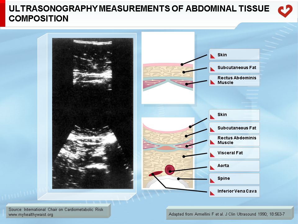
Similarly, ultrasonography can be used to estimate visceral fat by measuring visceral fat diameter (Figure 2). Visceral fat diameter is the distance between the abdominal muscle wall and the aorta, generally assessed around the level of the umbilicus or at L4-L5 [18,19]. However, it may be hard to measure visceral fat diameter if there are air-filled structures, such as the lungs or intestines, in the path of the ultrasound beam. Air has extremely low acoustic impedance, which means it reflects very little “echo” or signal back to the transducer. Consequently, if the aorta is located behind an air-filled intestine, ultrasound images may be obscured and the visceral diameter difficult to assess.
Ultrasonography-measured visceral fat thickness is generally a similar or stronger predictor of CT-measured visceral fat compared to anthropometric measures such as body mass index, waist circumference, waist-to-hip ratio, sagittal diameter, or skinfolds [19-23], or other clinical measures such as bioelectrical impedance analysis and DEXA [20]. Furthermore, ultrasonography is a better measure of changes in visceral fat compared to waist-to-hip ratio [24]. It is unclear whether ultrasonography is a better predictor of changes in visceral fat compared to waist circumference.
There is evidence that ultrasonography-measured visceral fat is a stronger predictor of health risk than simple anthropometric measures. Though some have reported that ultrasonography-measured visceral fat is a weaker predictor of health risk compared to waist circumference or DEXA [25], others have suggested that ultrasonography is a better measure of cardiovascular risk compared to waist circumference [18,21] and sagittal diameter [18]. Some have also reported that ultrasonography-measured visceral fat may be comparable to CT-measured visceral fat in predicting alterations in glucose metabolism and fasting lipid levels [26-28]. However, few studies have compared the utility of ultrasonography- and CT-measured visceral fat in predicting health risk, and their findings should be verified with further research.
To date, there is limited research on the usefulness of ultrasonography in assessing health risk compared to CT-measured visceral adiposity or anthropometric tools. Currently, ultrasonography appears to be a potentially useful clinical tool for assessing visceral fat and obesity-related health risk. However, further research is needed to confirm these findings.
References
-
Heymsfield SB, Lohman TG, Wang Z and Going SB. Human Body Composition. Human Kinetics Press: Champaign, IL , 2005.
PubMed ID:
-
Bray GA, Bouchard C and James WPT. Handbook of Obesity. Marcel Dekker, Inc.: New York, NY, 1998.
PubMed ID:
-
Brownbill RA and Ilich JZ. Measuring body composition in overweight individuals by dual energy x-ray absorptiometry. BMC Med Imaging 2005; 5: 1.
PubMed ID: 15748279
-
Kim J, Wang Z, Heymsfield SB, et al. Total-body skeletal muscle mass: estimation by a new dual-energy X-ray absorptiometry method. Am J Clin Nutr 2002; 76: 378-83.
PubMed ID: 12145010
-
Genton L, Hans D, Kyle UG, et al. Dual-energy X-ray absorptiometry and body composition: differences between devices and comparison with reference methods. Nutrition 2002; 18: 66-70.
PubMed ID: 11827768
-
Visser M, Fuerst T, Lang T, et al. Validity of fan-beam dual-energy X-ray absorptiometry for measuring fat-free mass and leg muscle mass. Health, Aging, and Body Composition Study–Dual-Energy X-ray Absorptiometry and Body Composition Working Group. J Appl Physiol 1999; 87: 1513-20.
PubMed ID: 10517786
-
Tylavsky F, Lohman T, Blunt BA, et al. QDR 4500A DXA overestimates fat-free mass compared with criterion methods. J Appl Physiol 2003; 94: 959-65.
PubMed ID: 12433854
-
Wang W, Wang Z, Faith MS, et al. Regional skeletal muscle measurement: evaluation of new dual-energy X-ray absorptiometry model. J Appl Physiol 1999; 87: 1163-71.
PubMed ID: 10484591
-
Salamone LM, Fuerst T, Visser M, et al. Measurement of fat mass using DEXA: a validation study in elderly adults. J Appl Physiol 2000; 89: 345-52.
PubMed ID: 10904070
-
Levine JA, Abboud L, Barry M, et al. Measuring leg muscle and fat mass in humans: comparison of CT and dual-energy X-ray absorptiometry. J Appl Physiol 2000; 88: 452-6.
PubMed ID: 10658010
-
Neeland IJ, Grundy SM, Li X, et al. Comparison of visceral fat mass measurement by dual-X-ray absorptiometry and magnetic resonance imaging in a multiethnic cohort: the Dallas Heart Study. Nutr Diabetes 2016; 6: e221.
PubMed ID: 27428873
-
Kaul S, Rothney MP, Peters DM, et al. Dual-energy X-ray absorptiometry for quantification of visceral fat. Obesity (Silver Spring) 2012; 20: 1313–1318.
PubMed ID: 22282048
-
Micklesfield LK, Goedecke JH, Punyanitya M, et al. Dual-energy X-ray performs as well as clinical computed tomography for the measurement of visceral fat. Obesity (Silver Spring) 2012; 20: 1109-1114.
PubMed ID: 22240726
-
Fourman LT, Kileel EM, Hubbard J, et al. Comparison of visceral fat measurement by dual-energy X-ray absorptiometry to computed tomography in HIV and non-HIV. Nutr Diabetes 2019; 9: 6.
PubMed ID: 30804324
-
Bellisari A, Roche AF and Siervogel RM. Reliability of B-mode ultrasonic measurements of subcutaneous adipose tissue and intra-abdominal depth: comparisons with skinfold thicknesses. Int J Obes Relat Metab Disord 1993; 17: 475-80.
PubMed ID: 8401751
-
Abe T, Tanaka F, Kawakami Y, et al. Total and segmental subcutaneous adipose tissue volume measured by ultrasound. Med Sci Sports Exerc 1996; 28: 908-12.
PubMed ID: 8832546
-
Abe T, Kondo M, Kawakami Y, et al. Prediction equations for body composition of Japanese adults by B-mode ultrasound. Am J Human Biol 1994; 6: 161-70.
PubMed ID: 28548275
-
Leite CC, Wajchenberg BL, Radominski R, et al. Intra-abdominal thickness by ultrasonography to predict risk factors for cardiovascular disease and its correlation with anthropometric measurements. Metabolism 2002; 51: 1034-40.
PubMed ID: 12145778
-
Tornaghi G, Raiteri R, Pozzato C, et al. Anthropometric or ultrasonic measurements in assessment of visceral fat? A comparative study. Int J Obes Relat Metab Disord 1994; 18: 771-5.
PubMed ID: 7866479
-
Kim SK, Kim HJ, Hur KY, et al. Visceral fat thickness measured by ultrasonography can estimate not only visceral obesity but also risks of cardiovascular and metabolic diseases. Am J Clin Nutr 2004; 79: 593-9.
PubMed ID: 15051602
-
Armellini F, Zamboni M, Rigo L, et al. The contribution of sonography to the measurement of intra-abdominal fat. J Clin Ultrasound 1990; 18: 563-7.
PubMed ID: 2170455
-
Ribeiro-Filho FF, Faria AN, Azjen S, et al. Methods of estimation of visceral fat: advantages of ultrasonography. Obes Res 2003; 11: 1488-94.
PubMed ID: 14694213
-
Armellini F, Zamboni M, Robbi R, et al. Total and intra-abdominal fat measurements by ultrasound and computerized tomography. Int J Obes Relat Metab Disord 1993; 17: 209-14.
PubMed ID: 8387970
-
Armellini F, Zamboni M, Rigo L, et al. Sonography detection of small intra-abdominal fat variations. Int J Obes 1991; 15: 847-52.
PubMed ID: 1794927
-
dos Santos RE, Aldrighi JM, Lanz JR, et al. Relationship of body fat distribution by waist circumference, dual-energy X-ray absorptiometry and ultrasonography to insulin resistance by homeostasis model assessment and lipid profile in obese and non-obese postmenopausal women. Gynecol Endocrinol 2005; 21: 295-301.
PubMed ID: 16373250
-
Ribeiro-Filho FF, Faria AN, Kohlmann O, Jr., et al. Ultrasonography for the evaluation of visceral fat and cardiovascular risk. Hypertension 2001; 38: 713-7.
PubMed ID: 11566963
-
Armellini F, Zamboni M, Harris T, et al. Sagittal diameter minus subcutaneous thickness. An easy-to-obtain parameter that improves visceral fat prediction. Obes Res 1997; 5: 315-20.
PubMed ID: 9285837
-
Liu KH, Chan YL, Chan WB, et al. Sonographic measurement of mesenteric fat thickness is a good correlate with cardiovascular risk factors: comparison with subcutaneous and preperitoneal fat thickness, magnetic resonance imaging and anthropometric indexes. Int J Obes Relat Metab Disord 2003; 27: 1267-73.
PubMed ID: 14513076
 CLOSE
CLOSE
 CLOSE
CLOSE
 CLOSE
CLOSE
Brownbill RA and Ilich JZ. Measuring body composition in overweight individuals by dual energy x-ray absorptiometry. BMC Med Imaging 2005; 5: 1.
PubMed ID: 15748279 CLOSE
CLOSE
Kim J, Wang Z, Heymsfield SB, et al. Total-body skeletal muscle mass: estimation by a new dual-energy X-ray absorptiometry method. Am J Clin Nutr 2002; 76: 378-83.
PubMed ID: 12145010 CLOSE
CLOSE
Genton L, Hans D, Kyle UG, et al. Dual-energy X-ray absorptiometry and body composition: differences between devices and comparison with reference methods. Nutrition 2002; 18: 66-70.
PubMed ID: 11827768 CLOSE
CLOSE
Visser M, Fuerst T, Lang T, et al. Validity of fan-beam dual-energy X-ray absorptiometry for measuring fat-free mass and leg muscle mass. Health, Aging, and Body Composition Study–Dual-Energy X-ray Absorptiometry and Body Composition Working Group. J Appl Physiol 1999; 87: 1513-20.
PubMed ID: 10517786 CLOSE
CLOSE
Tylavsky F, Lohman T, Blunt BA, et al. QDR 4500A DXA overestimates fat-free mass compared with criterion methods. J Appl Physiol 2003; 94: 959-65.
PubMed ID: 12433854 CLOSE
CLOSE
Wang W, Wang Z, Faith MS, et al. Regional skeletal muscle measurement: evaluation of new dual-energy X-ray absorptiometry model. J Appl Physiol 1999; 87: 1163-71.
PubMed ID: 10484591 CLOSE
CLOSE
Salamone LM, Fuerst T, Visser M, et al. Measurement of fat mass using DEXA: a validation study in elderly adults. J Appl Physiol 2000; 89: 345-52.
PubMed ID: 10904070 CLOSE
CLOSE
Levine JA, Abboud L, Barry M, et al. Measuring leg muscle and fat mass in humans: comparison of CT and dual-energy X-ray absorptiometry. J Appl Physiol 2000; 88: 452-6.
PubMed ID: 10658010 CLOSE
CLOSE
Neeland IJ, Grundy SM, Li X, et al. Comparison of visceral fat mass measurement by dual-X-ray absorptiometry and magnetic resonance imaging in a multiethnic cohort: the Dallas Heart Study. Nutr Diabetes 2016; 6: e221.
PubMed ID: 27428873 CLOSE
CLOSE
Kaul S, Rothney MP, Peters DM, et al. Dual-energy X-ray absorptiometry for quantification of visceral fat. Obesity (Silver Spring) 2012; 20: 1313–1318.
PubMed ID: 22282048 CLOSE
CLOSE
Micklesfield LK, Goedecke JH, Punyanitya M, et al. Dual-energy X-ray performs as well as clinical computed tomography for the measurement of visceral fat. Obesity (Silver Spring) 2012; 20: 1109-1114.
PubMed ID: 22240726 CLOSE
CLOSE
Fourman LT, Kileel EM, Hubbard J, et al. Comparison of visceral fat measurement by dual-energy X-ray absorptiometry to computed tomography in HIV and non-HIV. Nutr Diabetes 2019; 9: 6.
PubMed ID: 30804324 CLOSE
CLOSE
Bellisari A, Roche AF and Siervogel RM. Reliability of B-mode ultrasonic measurements of subcutaneous adipose tissue and intra-abdominal depth: comparisons with skinfold thicknesses. Int J Obes Relat Metab Disord 1993; 17: 475-80.
PubMed ID: 8401751 CLOSE
CLOSE
Abe T, Tanaka F, Kawakami Y, et al. Total and segmental subcutaneous adipose tissue volume measured by ultrasound. Med Sci Sports Exerc 1996; 28: 908-12.
PubMed ID: 8832546 CLOSE
CLOSE
Abe T, Kondo M, Kawakami Y, et al. Prediction equations for body composition of Japanese adults by B-mode ultrasound. Am J Human Biol 1994; 6: 161-70.
PubMed ID: 28548275 CLOSE
CLOSE
Leite CC, Wajchenberg BL, Radominski R, et al. Intra-abdominal thickness by ultrasonography to predict risk factors for cardiovascular disease and its correlation with anthropometric measurements. Metabolism 2002; 51: 1034-40.
PubMed ID: 12145778 CLOSE
CLOSE
Tornaghi G, Raiteri R, Pozzato C, et al. Anthropometric or ultrasonic measurements in assessment of visceral fat? A comparative study. Int J Obes Relat Metab Disord 1994; 18: 771-5.
PubMed ID: 7866479 CLOSE
CLOSE
Kim SK, Kim HJ, Hur KY, et al. Visceral fat thickness measured by ultrasonography can estimate not only visceral obesity but also risks of cardiovascular and metabolic diseases. Am J Clin Nutr 2004; 79: 593-9.
PubMed ID: 15051602 CLOSE
CLOSE
Armellini F, Zamboni M, Rigo L, et al. The contribution of sonography to the measurement of intra-abdominal fat. J Clin Ultrasound 1990; 18: 563-7.
PubMed ID: 2170455 CLOSE
CLOSE
Ribeiro-Filho FF, Faria AN, Azjen S, et al. Methods of estimation of visceral fat: advantages of ultrasonography. Obes Res 2003; 11: 1488-94.
PubMed ID: 14694213 CLOSE
CLOSE
Armellini F, Zamboni M, Robbi R, et al. Total and intra-abdominal fat measurements by ultrasound and computerized tomography. Int J Obes Relat Metab Disord 1993; 17: 209-14.
PubMed ID: 8387970 CLOSE
CLOSE
Armellini F, Zamboni M, Rigo L, et al. Sonography detection of small intra-abdominal fat variations. Int J Obes 1991; 15: 847-52.
PubMed ID: 1794927 CLOSE
CLOSE
dos Santos RE, Aldrighi JM, Lanz JR, et al. Relationship of body fat distribution by waist circumference, dual-energy X-ray absorptiometry and ultrasonography to insulin resistance by homeostasis model assessment and lipid profile in obese and non-obese postmenopausal women. Gynecol Endocrinol 2005; 21: 295-301.
PubMed ID: 16373250 CLOSE
CLOSE
Ribeiro-Filho FF, Faria AN, Kohlmann O, Jr., et al. Ultrasonography for the evaluation of visceral fat and cardiovascular risk. Hypertension 2001; 38: 713-7.
PubMed ID: 11566963 CLOSE
CLOSE
Armellini F, Zamboni M, Harris T, et al. Sagittal diameter minus subcutaneous thickness. An easy-to-obtain parameter that improves visceral fat prediction. Obes Res 1997; 5: 315-20.
PubMed ID: 9285837 CLOSE
CLOSE
Liu KH, Chan YL, Chan WB, et al. Sonographic measurement of mesenteric fat thickness is a good correlate with cardiovascular risk factors: comparison with subcutaneous and preperitoneal fat thickness, magnetic resonance imaging and anthropometric indexes. Int J Obes Relat Metab Disord 2003; 27: 1267-73.
PubMed ID: 14513076
